


A 2007 clinical report by the Department of Plastic Surgery at the University of Verona, Italy — When autologous fat (including SVF) was transplanted into tissue hardened by radiation therapy, tissue hydration and angiogenesis improved, and both symptoms and cosmetic results were enhanced. Following this paper, the paradigm of fat grafting changed.
SC301 Clinic · Stem Cell Research Series 4th Week of June · Single Paper Based · Direct Transplantation of Pure SVF (Lipoaspirate)
Original paper information:
Rigotti G, Marchi A, Galiè M, Baroni G, Benati D, Krampera M, Pasini A, Sbarbati A.
“Clinical treatment of radiotherapy tissue damage by lipoaspirate transplant: a healing process mediated by adipose-derived adult stem cells”
Plastic and Reconstructive Surgery. 2007;119(5):1409–1422. doi: 10.1097/01.prs.0000256047.47909.71. PMID: 17415234.
Affiliation: Department of Plastic Surgery, University of Verona, Italy / Joint research team including the Department of Neuroscience, Morphology, and Exercise Science
2007, a turning point in regenerative medicine — things are different before and after this paper.
Fat grafting (lipofilling) has long been perceived as a simple "volume-filling" procedure. The principle is that when fat is harvested and transplanted to the necessary areas, volume is maintained for as long as that fat survives.
However, this paper published by the Rigotti G research team in 2007 completely changed that perception.
The research team transplanted autologous fat (lipospirate) into severely damaged tissue after radiation therapy, going beyond simple volume restoration. The phenomenon in which the tissue itself regenerates and new blood vessels are createdThis was systematically reported for the first time in clinical practice. This paper argues that fat grafting is not a 'simple volume filler' Stem cell-mediated regenerative therapyIt was a pioneering study that proved that it could be, and subsequently changed the course of regenerative medicine research worldwide.
800+
Citation count of this paper (as of 2024)
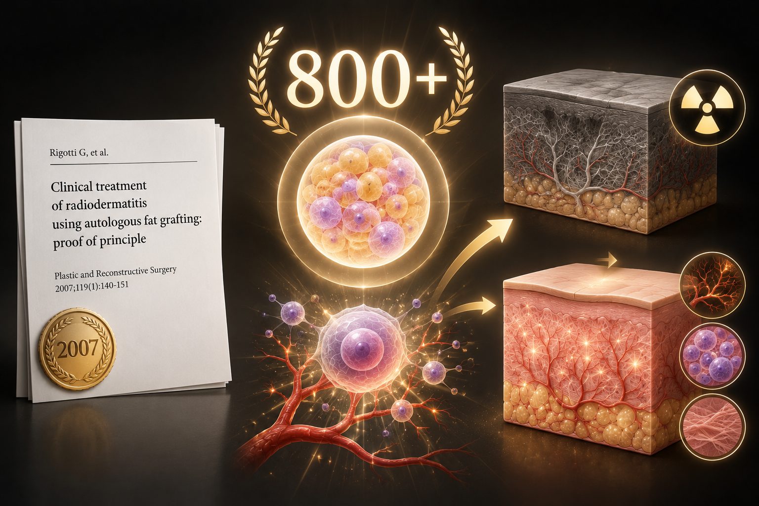
As one of the most important early clinical evidences in the field of radiation tissue reconstruction
It is continuously cited in plastic surgery and regenerative medicine research worldwide.
Key finding of this paper: When fat (including SVF) is transplanted into radiation-damaged tissue, adipose-derived stem cells (ADSCs) promote angiogenesis, reduce tissue inflammation, and revitalize fibrotic tissue — this was the first time this was clinically proven.
What happens to tissues after radiation therapy
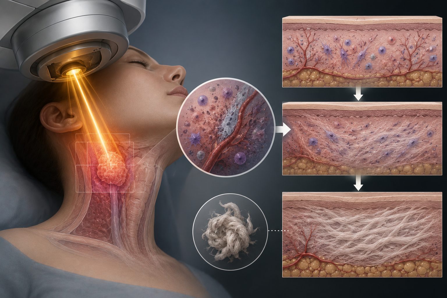
While radiation therapy is a highly effective weapon in cancer treatment, it leaves irreversible damage to normal tissues surrounding the target site. Radiation-Induced Fibrosis (RIF)It is called.
1
Immediately after radiation exposure
Damage to vascular endothelial cells → Blocking of blood supply → Beginning of oxygen and nutrient deficiency in tissues
2
months to years have passed
Persistent chronic inflammation → TGF-β overexpression → Excessive collagen accumulation → Tissue fibrosis (hardening)
3
progression of fibrosis
Hardening of the skin, subcutaneous tissue, and muscles → Occurrence of pain, stiffness, deformation, and skin ulcers
XNUMX
Limitations of existing treatments
Drug and physical therapy only alleviate symptoms. In severe cases, reconstructive surgery is necessary, but the results are poor. Prior to this paper, RIF was considered virtually 'irreversible'.
Typical examples include the hardening of the skin and subcutaneous tissue to leave scars after radiation therapy for breast cancer, or the stiffening of the face after radiation therapy for head and neck cancer. This is precisely what Rigotti's research team focused on. Can RIF, which was considered 'irreversible,' be reversed through fat grafting?It was.
How the research was conducted — Clinical research at the University of Verona, Italy
Rigotomy — A proprietary technique developed by this research team
What is Rigotomy?
The technique developed by Dr. Rigotti's team is more than simple fat grafting. Due to the dense connective tissue in fibrotic tissue, there is no space for fat to enter, and even if it is injected, engraftment is difficult. To solve this, the research team Percutaneous FasciotomyPerform this first.
In other words, it is a method in which hardened fibrous tissue is finely separated and broken down using a special needle without incising the skin, and then fat (including SVF) is injected into the resulting empty space.
This technique is later RigotomyIt is called by this name and has established itself as a standard approach to maximize the effectiveness of fat grafting in 'areas with poor grafting environments,' such as radiation damage, burns, and severe scars.
Why Fat Grafting Restores Radiation Damage — The Role of SVF

What the research team focused on was what is contained within the fat stromal vascular fraction (SVF)Lipoaspirates contain not only adipocytes but also SVF cells with mesenchymal stem cell characteristics. These SVF cells function in radiation-damaged tissue as follows:
Mesenchymal stem cells (MSC)
Promotion of angiogenesis, inhibition of inflammation, and secretion of signals for fibrosis reversal. The key protagonists of this paper
Vascular endothelial cells
Directly participates in the repair of radiation-damaged vascular networks. The core of tissue reperfusion.
prefat cells
Differentiation into new adipocytes contributes to the restoration of normal soft tissue structure in fibrotic areas.
Immunomodulatory cells
Suppression of chronic inflammation persisting after radiation, creation of an environment favorable for tissue regeneration
Radiation Damage → Pathway of Tissue Change After SVF Transplantation

① Angiogenesis-promoting pathway
Radiation → Vascular destruction → Chronic ischemia/fibrosis → SVF transplantation → VEGF/HGF secretion → Renal angiogenesis → Tissue hydration/reperfusion
② Antifibrotic pathway
Radiation → TGF-β Overexpression → Collagen Overaccumulation and Fibrosis → SVF → Secretion of Antifibrotic Factors → Reversal of Fibrosis → Restoration of Tissue Flexibility
③ Anti-inflammatory pathway
Radiation → Induction of chronic inflammation → Persistent tissue damage → SVF → IL-10, immunomodulatory factor → Suppression of inflammation → Creation of a regenerative environment
What happened after the transplant — both symptoms and tissue changed
Pre-transplant — Condition of radiation-damaged tissue
Hard, painful, and dry tissue
● Hardness due to fibrosis of skin and subcutaneous tissue
● Tissue drying and loss of elasticity due to blood vessel destruction
● Persistent pain and stiffness
● Some patients have accompanying skin ulcers
● Cosmetic deformity, depression, asymmetry
Post-transplant — Changes after SVF transplantation
Hydrated and regenerated tissue
● Improvement of tissue hydration
● Restoration of blood flow through angiogenesis
● Improvement of symptoms (pain, stiffness)
● Skin ulcer recovery
● Improvement of cosmetic results
1
Simultaneous confirmation of symptomatic and cosmetic improvement — in all 20 patients
After autologous fat grafting including SVF in all 20 patients with radiation fibrosis Simultaneous confirmation of symptomatic and aesthetic improvementThat is it. It is important to note that not only has the outward appearance improved, but the symptoms the patient actually feels, such as pain and stiffness, have also improved.
2
Improved tissue hydration — Hardened tissue became moist
dry and parched tissues due to radiation damage Hydration improvedThat is it. The research team interpreted this as the result of increased blood supply caused by the formation of a new vascular network. The fact that the tissue is becoming moist again is an important signal that living regeneration is taking place.
3
Angiogenesis — The destroyed vascular network was recreated
In tissue after transplantation Neo-angiogenesis confirmedThat is it. The vascular network destroyed by radiation was regenerated through the cooperation of vascular endothelial cells and MSCs within the SVF. This was the most shocking discovery delivered by this paper to the academic community—it was direct evidence that radiation-damaged tissue can regenerate rather than being 'irreversible'.
XNUMX
Histological analysis — Confirmation of regeneration at the cellular level
Through clinical observation as well as tissue biopsies before and after transplantation Histological analysisChanges were also confirmed in this. The cells in SVF (lipospirates) Physical Phenotype and Immunophenotype of Mesenchymal Stem Cells (MSCs)It was confirmed that they possess [these elements] and suggested that they are mediators of angiogenesis and anti-inflammatory action.
What Has Changed Since This Paper — A Turning Point in Regenerative Medicine
Prior to the publication of this paper by Rigotti G's research team in 2007, radiation-induced fibrosis (RIF) was considered virtually "irreversible damage." While symptoms could be somewhat alleviated with medication or physical therapy, there was no concept that damaged tissue could fundamentally regenerate.
This paper overturned that perception. The research team went beyond simply stating that "fat grafting is effective," The effect is mediated by adipose stem cells (ADSC/MSC).It also presented the mechanistic basis. Since then, hundreds of follow-up studies have verified and expanded upon the findings of this paper, and lipofilling has newly established itself as a stem cell-mediated regenerative therapy rather than a simple cosmetic procedure.
The conclusion reached by the paper
Autologous lipospirate (including SVF) transplantation in all 20 patients with radiation-induced tissue injury (RIF) Symptomatic and cosmetic improvementIt elicited.
The key mechanism is lipospirate Adipose-derived adult stem cell (ADSC)-mediated promotion of angiogenesis and improvement of tissue hydrationThis healing process took place in a manner where MSCs supported angiogenesis and reduced tissue inflammation.
This paper is the first systematic report on the use of fat grafting in the clinical treatment of radiation tissue damage, The turning point that redefined fat grafting as 'stem cell-mediated regenerative therapy'It has become.
Stories from SC301 Clinic
The most important message of this paper is one — What was thought to be irreversible can be reversedThis is the first clinical demonstration that tissue hardened by radiation regenerates by forming new blood vessels and retaining moisture through the action of stem cells in the fat.
Today, treatments utilizing SVF and fat grafting for scar treatment, radiation damage reconstruction, and facial reconstruction are being performed worldwide, all built upon the foundation laid by this paper.
SC301 Clinic will honestly deliver the scientific foundation of stem cell (SVF) therapy, ranging from evidence-based papers that changed the history of regenerative medicine to the latest clinical trials.
Questions (Q&A) regarding this study
Q1. Why does the area treated with radiation therapy harden like that?
A. This is due to a process called radiation-induced fibrosis (RIF). While radiation effectively kills cancer cells, it also leaves damage to normal tissues surrounding the target. First, immediately after radiation exposure, vascular endothelial cells are damaged, cutting off blood supply and leaving the tissues deprived of oxygen and nutrients. Subsequently, as chronic inflammation persists over several months to years, a factor called TGF-β is overexpressed, and collagen accumulates excessively, causing the tissues to harden. As the condition progresses, fibrosis spreads to the skin, subcutaneous tissue, and muscles, leading to pain, stiffness, deformation, and, in severe cases, even skin ulcers. Prior to the publication of this study in 2007, such hardened tissue was considered virtually 'irreversible,' meaning it could not be restored.
Q2. How can hardened tissue be regenerated when it is simply filled with fat?
A. That is precisely the core of this paper. This is because the study demonstrated that fat grafting is not merely about filling volume. The harvested fat (lipoaspirates) contains not only fat cells but also SVF (stromal vascular fraction) cells, which possess mesenchymal stem cell characteristics. These cells operate in damaged tissue through three pathways. First, they secrete factors such as VEGF and HGF to regenerate damaged vascular networks (anegiogenesis), thereby restoring blood flow and tissue hydration. Second, they secrete anti-fibrotic factors to restore flexibility to tissues hardened by over-accumulated collagen. Third, they suppress chronic inflammation using immunomodulatory factors such as IL-10, creating an environment favorable for regeneration. In other words, it does not simply fill space but changes the tissue environment itself at the cellular level.
Q3. How is 'Rigottomy' different from regular fat grafting?
A. Fibrotic tissue lacks the space for fat due to its dense connective tissue, and even if injected, it does not take root well. To solve this problem, Dr. Rigotti's team developed a technique called Rigotomi. This method involves first performing a percutaneous fasciotomy using a special needle to finely separate and deconstruct the hardened fibrous tissue without making a large incision in the skin, and then injecting fat containing SVF into the resulting empty space. Thanks to this technique, the effectiveness of fat grafting can be enhanced even in areas with poor graft environments, such as radiation damage, and it is now being applied as a standard procedure to other challenging areas, including burn scars, severe acne scars, and scar contractures.
Q4. What changes were actually observed in the patients?
A. In this study, both symptomatic and cosmetic improvements were simultaneously confirmed in all 20 patients who had developed fibrosis following breast cancer radiation therapy. Importantly, the improvement was not limited to outward appearance. The hydration (moisture content) of the hardened and dried-out tissues improved, and symptoms actually experienced by the patients, such as pain and stiffness, also improved; furthermore, skin ulcers in some patients healed. In addition, the study did not stop at clinical observation but confirmed changes through histological analysis using tissue biopsies performed before and after transplantation, directly demonstrating the formation of new blood vessels to replace the destroyed vascular network. This discovery came as a major shock to the academic community, as it presented direct evidence that tissue, previously considered irreversible, can regenerate.
Q5. Can the results of this study be directly applied to facial radiation injury?
A. Caution is required when making direct generalizations. This study was a relatively small-scale, single-group observational study involving 20 participants without a randomized control group, and the subjects primarily focused on fibrosis in the chest and breast areas following radiation therapy for breast cancer. Therefore, additional evidence is needed to immediately guarantee identical results when applied to other body parts, such as facial injuries following radiation therapy for head and neck cancer. However, since the fundamental mechanisms of tissue regeneration—namely angiogenesis, anti-fibrosis, and anti-inflammation—operate identically regardless of the body part, the findings of this study provide a theoretical foundation for the reconstruction of facial radiation injuries. In fact, hundreds of follow-up studies have verified and expanded upon these findings since this paper, broadening their application to various body parts. In other words, it is accurate to understand this as a solid starting point rather than an "established standard."

Frequently Asked Questions
What happens to tissues after radiation therapy
While radiation therapy is a highly effective weapon in cancer treatment, it leaves irreversible damage to normal tissues surrounding the target site. This is called Radiation-Induced Fibrosis (RIF). 1 Damage to vascular endothelial cells immediately after radiation exposure → Blockage of blood supply → Beginning of oxygen and nutrient deficiency in tissues


