A research team at Alexandria Medical University in Egypt transplanted SVF-enhanced fat and regular fat into the temples of the same patient and precisely measured volume durability after 6 months using a bio-ultraviolet microscope (UBM).
SC301 Clinic · Stem Cell Research Series 3th Week of June · Single Paper Based · Pure SVF Direct Transplantation
📄 Original paper information
Roshdy OH, Abdallah WI, Farid CI, Mehanna RA, Bayoumi NH, Ismail AI.
"Stromal vascular fraction improves the durability of autologous fat temple augmentation — a split-face randomized study using ultrasound biomicroscopy"
Journal of Plastic, Reconstructive & Aesthetic Surgery (JPRAS). 2022;75(6):1870–1877.
doi: 10.1016/j.bjps.2022.01.003. PMID: 35125305. ClinicalTrials.gov NCT03965936.
Affiliation: Department of Plastic Surgery and Medical Physiology, Faculty of Medicine, University of Alexandria (Egypt)
When discussing facial aging, wrinkles around the eyes or nasolabial folds are commonly mentioned first.
However, one of the areas that plastic surgeons cite as a key indicator of aging is... Temple.
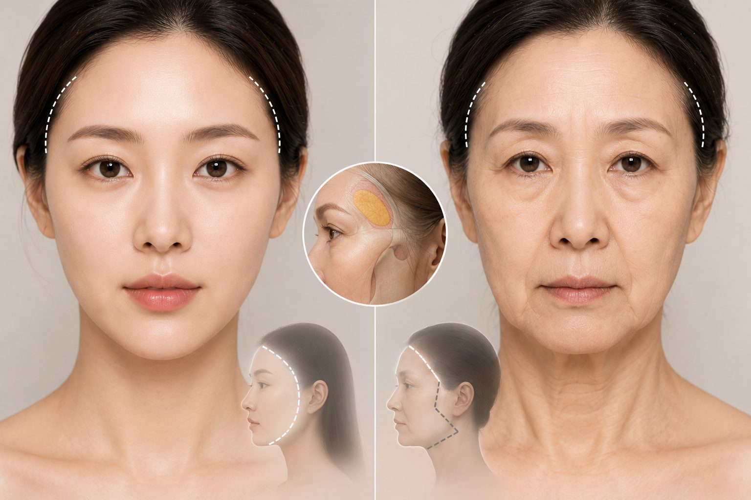
The temples are the excess area between the forehead and the cheeks; they maintain a gentle bulge in youth but gradually sink as one ages.
If the temples are sunken, the forehead looks wider, and the face turns into an inverted triangle 'elderly silhouette'.
You can add volume with fat grafting (lipofilling), but the biggest problem is How long does the transplanted fat survive?.
This is the core question that this study sought to answer.
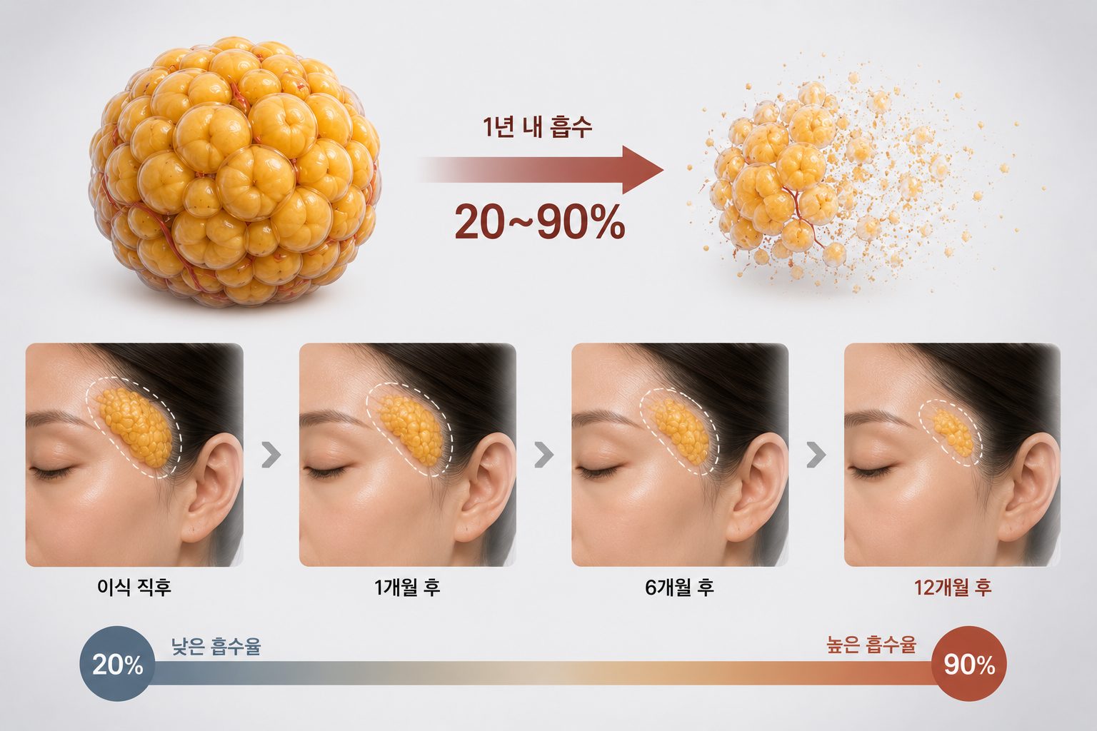
Autologous fat grafting is an excellent method for creating natural volume without worrying about immune rejection, but it has limitations that trouble both doctors and patients. Of the transplanted fat 20–90% is absorbed and disappears in the first year.It is.
Because this variation is so large, even if the same doctor performs the procedure using the same technique, the results vary significantly from patient to patient.
Especially The temple area is known to have a low fat survival rate due to its relatively poor blood supply..
This research team utilized SVF right here on the temple.
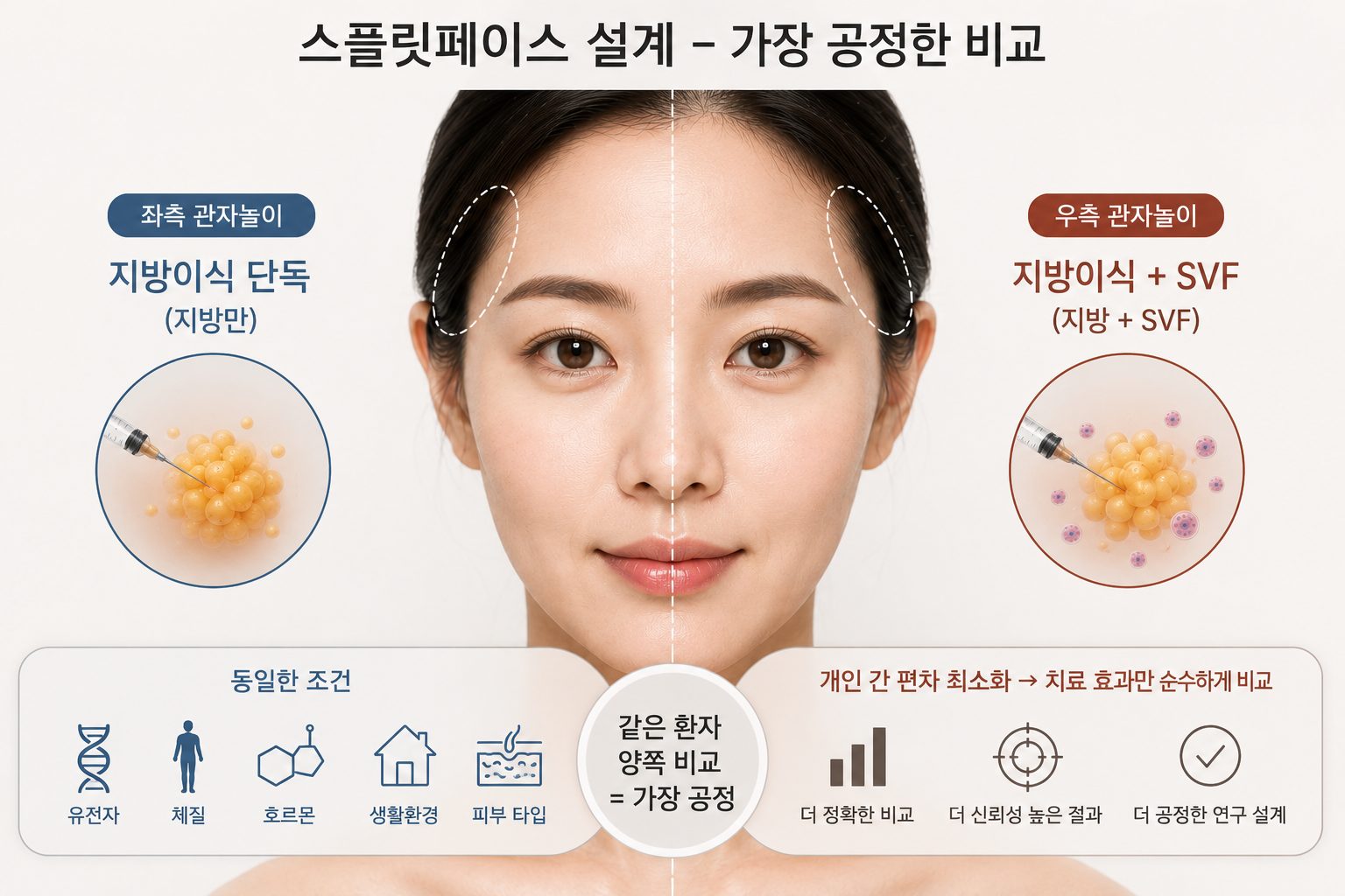
Why is Split-Face Design Powerful?
The split-face design, which applies different treatments to both sides of the same patient's face, allows for the comparison of two treatments under conditions where genetics, constitution, hormones, lifestyle, and skin type are completely identical.
Rather than studies comparing two different patient groups Minimizing inter-individual variation to purely compare treatment effectsYou can.
The SVF enhancement method used in this study is similar to the CAL concept of Dr. Yoshimura's team introduced earlier.
It is a method in which a portion of the fat is used for SVF extraction, and that SVF is mixed with the remaining fat for transplantation.
Fat harvesting → Separation of SVF from a portion → Mixing of transplantable fat + SVF → SVF-enhanced fat temple transplant
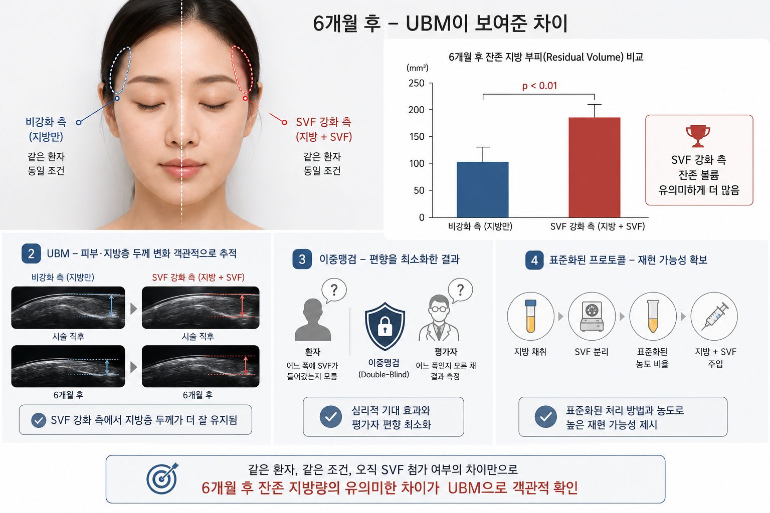
The distinguishing feature of this study is Standardized SVF concentration ratio and treatment protocolThe point is that it used
To address criticism that previous studies yielded inconsistent results due to the use of various concentration ratios and treatment methods, the research team designed a reproducible standard protocol.
The temples have an anatomical characteristic of relatively insufficient blood supply.
For transplanted fat cells to survive, a vascular network must form rapidly, but this process is slow in the temples.
The way SVF solves this problem is as follows.
Rapid angiogenesis
Vascular endothelial cells and ADSCs in SVFs accelerate angiogenesis immediately after transplantation through VEGF secretion.
Ischemic defense
Protective factors of ADSCs inhibit adipocyte apoptosis during the ischemic period prior to vascular network formation
Adipocyte differentiation
Preadipocytes differentiate into new adipocytes, contributing to the long-term maintenance of transplant volume.
Immunomodulation
Creating a favorable engraftment environment for transplanted fat by suppressing excessive inflammatory responses
The greatest strength of this study is Split-phase + Double-blind + Standardized Protocol + Objective Measurement (UBM)The point is that it simultaneously satisfied the four conditions.
This is an attempt to resolve the problem where SVF-enhanced fat grafting studies yielded inconsistent results due to the use of different methods and concentrations, and to prove the efficacy of SVF using a reproducible standard method.
Because the split-phase design completely controls for individual differences, it can demonstrate strong therapeutic effects even in small-scale studies.
The key to its reliability is that this study is a randomized double-blind study, not a simple case series or observational study.

In temporal fat grafting using a standardized SVF reinforcement protocol, the SVF reinforcement side After 6 months, the residual fat volume was statistically significantly higher compared to the non-reinforced side.
Ultrasound-based biomicroscopy (UBM) has been confirmed to be a useful objective tool for tracking changes in the thickness of the temporal skin and fat layers. These results support the existing hypothesis that SVF reinforcement improves the durability of temporal fat grafting, and Verification through additional large-scale, long-term studies is required.
Q&A regarding this study
Q1. How much higher is the engraftment rate when SVF is mixed in for temporal fat grafting?
A. In a split-face randomized double-blind study published by a research team at the Faculty of Medicine, Alexandria University in Egypt, residual volume was measured using an ultrasound biomicroscope six months after SVF-enhanced fat and standard fat were transplanted to the temples of the same patient. The results confirmed that the amount of residual fat remaining on the side with added SVF was statistically significantly greater than on the side without. The strength of this study lies in the comparison of both faces of the same patient. Since the study was conducted under conditions where genetics, constitution, hormones, and lifestyle were completely identical, differing only in whether SVF was added, it was possible to purely measure the effects of SVF while completely controlling for variables caused by individual differences. However, as the research team themselves acknowledged the limitations of a six-month follow-up period and the study being conducted on a single site, follow-up research involving long-term data of over 12 months and a wider area is currently required. SC301 has continuously reviewed these latest research trends and advanced clinical trials in a direction that increases the engraftment rate of SVF-based fat grafting, and the average engraftment rate of 77.48% officially reported in an SCI-level international academic journal is the result of this accumulated research.
Q2. Why is the survival rate of fat grafts in the temples lower than in other areas?
관자놀이는 해부학적으로 혈액 공급이 상대적으로 부족한 부위입니다. 이식된 지방세포가 새로운 환경에서 살아남으려면 주변에 혈관망이 빠르게 형성되어 산소와 영양 공급이 이루어져야 합니다. 그런데 관자놀이는 이 혈관 형성 과정이 다른 부위보다 느리기 때문에 이식 직후 지방세포가 영양 공급을 받지 못하는 허혈 기간이 길어지고, 그 사이에 지방세포가 사멸하는 비율이 높아집니다. 이것이 관자놀이 지방이식에서 생착률이 낮고 시간이 지나면서 볼륨이 빠르게 꺼진다는 경험담이 많은 이유입니다. SVF에 포함된 혈관내피세포와 지방유래 줄기세포는 VEGF라는 혈관 형성 촉진 인자를 분비해 이식 직후부터 혈관망 형성을 가속화합니다. 또한 혈관망이 완성되기 전 허혈 기간 동안 지방유래 줄기세포의 보호 인자가 지방세포 사멸을 억제하는 역할을 합니다. 관자놀이처럼 혈액 공급이 부족한 부위일수록 SVF 강화의 효과가 더 뚜렷하게 나타날 수 있는 이유가 바로 여기에 있습니다. SC301은 자체 줄기세포 생착 연구센터를 운영하며 이러한 생착 환경 개선에 대한 연구를 18년 이상 이어오고 있습니다.
Q3. Why is the split-phase study design important, and why is it more reliable than other studies?
The biggest obstacle when comparing two treatments in clinical studies is individual differences, such as varying genes, constitution, immune responses, and lifestyles. When comparing two different groups of patients, it is difficult to distinguish whether the treatment is actually more effective or if the patients in that group simply had a constitution that inherently resulted in a higher engraftment rate. The Split-Face design fundamentally resolves this problem. Because different treatments are applied simultaneously to both sides of the same patient's face, it allows for a comparison solely of differences in treatment methods under conditions where genes, constitution, hormones, immune responses, living environments, and skin types are completely identical. The recent University of Alexandria study further enhanced reliability by incorporating a double-blind design. Since the patient was unaware of which side the SVF was administered to, and evaluators measured ultrasound readings in a blind state, the possibility of psychological expectations or evaluator bias influencing the results was minimized. This is why this design is recognized as strong evidence, despite being a small-scale study. SC301 was able to publish its SVF-related research results in international SCI-indexed journals, including the Oxford Journal, precisely because it was underpinned by such rigorous research design and the accumulation of objective data.
Q4. Why is measuring fat engraftment using Ultrasonic Biomicroscopy (UBM) better than existing evaluation methods?
Until now, methods for evaluating the survival rate of fat grafts have primarily relied on comparisons of before-and-after photos, visual evaluations by physicians, or surveys of patients' subjective satisfaction. These methods are susceptible to unscientific variables, such as lighting angles, camera settings, evaluator subjectivity, and patient expectations. Ultrasonic Biomicroscopy (UBM) utilizes frequencies of 35–100 MHz—much higher than standard ultrasound—to measure skin and subcutaneous tissue with millimeter-level precision. By quantifying changes in the thickness and density of transplanted fat numerically, it allows for objective comparisons of the pre- and post-procedure states, enabling the capture of even minute, invisible changes in data. It was thanks to this objective measurement tool, UBM, that the University of Alexandria study was able to statistically demonstrate a significant difference in residual fat volume between the SVF-enhanced and non-enhanced sides. This is also why the reliability of this study is highly regarded, as it quantified differences that are difficult to capture through subjective evaluations or photo comparisons. This aligns with the direction in which SC301 has accumulated clinical evidence based on objective data in SVF survival research.
Q5. Can I expect the same effects on other facial areas besides the temples after receiving SVF reinforced fat grafting?
Because this study from the University of Alexandria focused on a single site—the temples—additional verification is required to apply the results directly to other facial areas. The research team itself acknowledges this as a limitation. However, the principle by which SVF enhances engraftment rates operates identically regardless of the site. The mechanism by which vascular endothelial cells and adipose-derived stem cells secrete VEGF to accelerate angiogenesis, inhibit adipocyte apoptosis during periods of ischemia, and differentiate preadipocytes into new adipocytes can be expected to be the same in fat grafting for other facial areas, such as the cheeks, forehead, nasolabial folds, and under-eye hollows. In particular, the effects of SVF enhancement are likely to be more pronounced in areas with relatively insufficient blood supply, such as the temples, or in areas with thin fat layers. SC301 has accumulated data on improving engraftment environments in various areas, including the temples, through over 18 years of research and clinical trials on SVF-based fat grafting across the entire face. If you are considering restoring volume to your entire face as well as your temples, we recommend that you check the specific direction tailored to the condition of each area through a 1:1 consultation with SC301.